头部背景图片

What Is The Principle Of SEM?

Scanning Electron Microscopy (SEM) is based on the principle of using a focused beam of high-energy electrons to probe the surface of a sample and produce a high-resolution detailed image.


Electron Source: SEM works by using an electron source, typically a heated tungsten filament or a field emission gun, to produce a beam of electrons.


Electron Beam Generation: The electron source emits electrons, which are accelerated to high energies by an electric field. The electrons are focused into a narrow beam using electromagnetic lenses.


Sample Interaction: The primary electron beam is directed onto the sample's surface. When the beam interacts with the sample, several types of interactions occur, including scattering, absorption, and emission of secondary electrons.


Scattering: The primary electrons may undergo elastic or inelastic scattering while interacting with the atoms in the sample. Elastic scattering results in a change in direction of the electron beam, while inelastic scattering leads to energy loss due to interactions with the sample's atoms.


Secondary Electron Emission: Some of the primary electrons knock off secondary electrons from the surface of the sample through inelastic scattering. These secondary electrons carry information about the sample's topography and composition.


Signal Detection: The emitted secondary electrons, along with other signals such as backscattered electrons and characteristic X-ray emissions, are detected using various detectors. Some common detectors in SEM are the Everhart-Thornley detector for secondary electrons and detectors for backscattered electrons or X-rays generated by the sample.


Image Formation: The detected signals are then amplified and processed to form an image. The signal intensity is typically converted into a grayscale or false-color representation, allowing the visualization of surface features and details.


Scanning: To generate a complete image, the electron beam is systematically scanned across the surface of the sample in a raster pattern. The intensity of the detected signals at each point is recorded, allowing the construction of a high-resolution image.


Image Display and Analysis: The final reconstructed image is displayed on a monitor or recorded for further analysis. SEM images can be used to examine the microstructure, morphology, elemental composition, and surface characteristics of a wide range of materials.


In short, scanning electron microscopy utilizes the interaction of a focused, high-energy electron beam with a sample to generate detailed images. By analyzing the signals emitted by the sample, SEM provides valuable information about the surface topography, morphology, and composition of the sample at high resolution. It is widely used in a variety of scientific and industrial applications for research, quality control, and materials characterization.


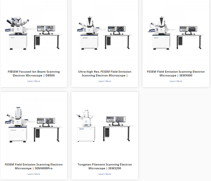
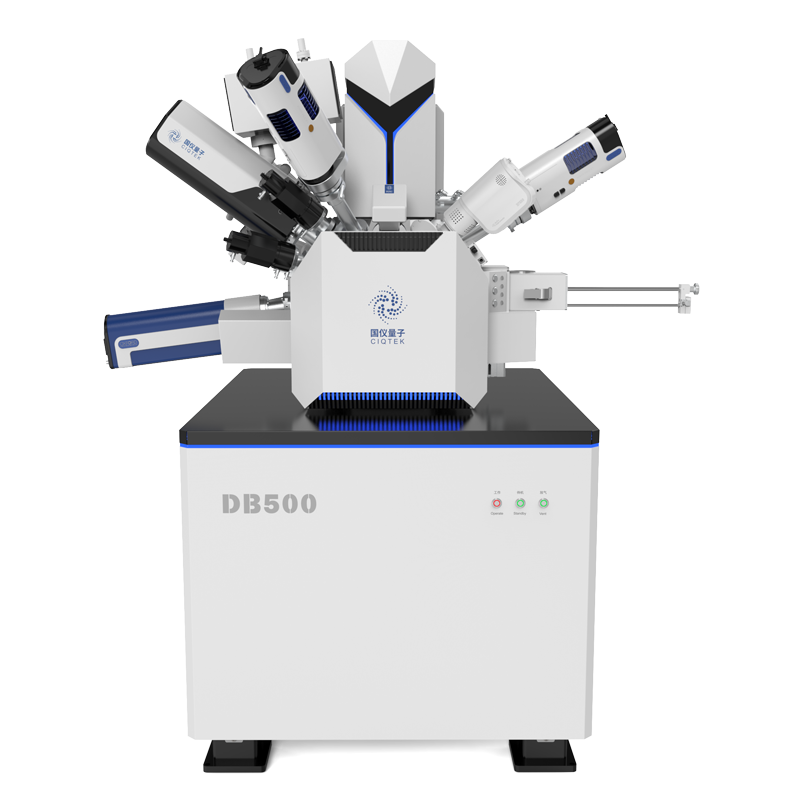
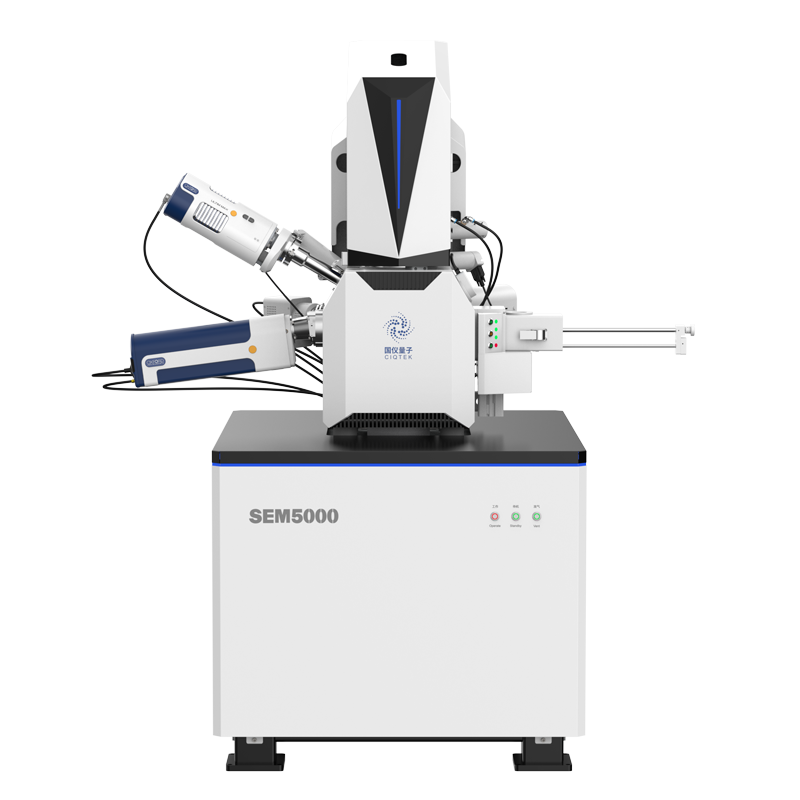
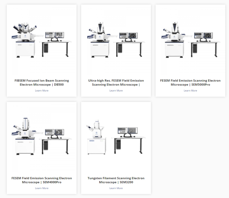
CIQTEK offers various high-quality SEM equipment for researchers and industrial users.
 

Scanning Electron Microscope Detectors

The Scanning Electron Microscope (SEM) is a powerful tool for various applications in material sciences, life sciences, and other fields. Different kinds of detectors have been developed to obtain more information and improve the performance of SEM. The following are a few common types of SEM detectors:


Backscattered Electron Detector (BSE): BSE detectors are used to detect electrons scattered by the nuclei of atoms and high atomic number elements within a substance.BSE detectors provide high-contrast images that can be used for compositional analysis of materials and characterization of microstructures.


Secondary Electron Detector (SE): The SE detector is used to detect secondary electrons on the surface of a material excited by a scanning electron beam. Since the surface morphology and material composition influence the secondary electrons, the SE detector provides a high-resolution image of the surface topography.


Transmission Electron Detector (TED): TED detectors are used to detect transmission electrons that pass through a thin sheet of material and are focused on the detector. TED detectors are suitable for high-resolution compositional and structural analyses of materials, e.g., atomic-level characterization of nanoparticles.


Energy Dispersive X-ray Spectroscopy (EDS): EDS detectors are used to analyze the elemental composition of materials. When a scanning electron beam interacts with a sample, characteristic X-rays are produced, and the EDS detector collects and measures the energy spectrum of these X-rays to determine the chemical composition and elemental distribution of the material.


Retarding Field Detector (RFD): The RFD detector is used to measure the charge carried by electrons generated on the surface of the sample. This type of detector is very useful for studying the electrical conductivity and surface charge properties of materials.


These are just some of the common SEM detectors; in fact, there are many other types of detectors, each with different advantages and applications. The selection of the appropriate detector depends on the objectives of the study and the information to be obtained.


CIQTEK's self-developed SEM offers a wide range of detectors to choose from, such as BSED, STEM, EDS, EDX, EBSD, In-lens, ETD, etc. 


Why ESR Is Also Known As EPR?

ESR (Electron Spin Resonance) and EPR (Electron Paramagnetic Resonance) are used interchangeably to describe the same spectroscopic technique. The reason for the two different names can be traced to the historical development of the field and some of the interesting stories surrounding it.


Originally, the technique was called ESR, or electron spin resonance. It was discovered in the mid-20th century by physicists studying the behavior of electrons in magnetic fields. They observed that certain materials absorbed energy at specific frequencies when exposed to strong magnetic fields and subjected to electromagnetic radiation. This absorption is due to the electron spin states flipping in the magnetic field, resulting in resonance.


As the field grew, researchers began to study the spectra of paramagnetic species - atoms, molecules, or ions with unpaired electrons. They realized that ESR techniques could be used to study a wider range of systems than just electrons. As a result, the term EPR (Electron Paramagnetic Resonance) was coined to encompass a broader range of applications.


The transition from ESR to EPR did not happen overnight or with universal adoption. Different research groups and scientific communities preferred to use either term. For example, the term ESR was widely used in the United States, while EPR became popular in Europe. This difference in naming conventions continued for some time until the two terms gradually became synonymous. Today, the two terms are widely accepted and used interchangeably to describe the same spectroscopic techniques.


The dual naming of ESR and EPR provides insight into the historical development of the field and how scientific terminology has evolved and adapted over time. It also serves as a reminder that in science, different cultures and communities may adopt different names for the same concept, and these differences may persist until a consensus is reached.


EPR spectroscopy has applications in a variety of fields including chemistry, physics, biochemistry, materials science, and medicine. It has been used to study the structure of metalloproteins, the mechanism of enzyme reactions, the properties of organic radicals, the behavior of transition metal complexes, and defects in semiconductors, as well as many other fascinating areas of research. Its ability to probe electron spin makes it an invaluable tool for understanding the fundamental properties of paramagnetic systems and exploring their role in a variety of physical and chemical processes.

>> Check more about CIQTEK EPR spectroscopy.

CIQTEK EPR spectroscopy

How To Find ESR Spectroscopy At Best Price

To find the best price on an Electron Spin Resonance (ESR) spectroscopy, you can follow the steps below:


1. Explore search platforms

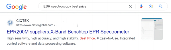
The most direct way is to search for “Electron Spin Resonance (ESR) spectroscopy best price” on major search engines like Google. Use words like cheap, sale, affordable, etc. to describe your needs, and it will be straightforward to find the right EPR spectroscopy at a recognized price, such as the one shown below:

best price Electron Spin Resonance spectroscopy


2. Explore online B2B marketplaces

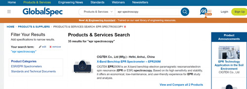
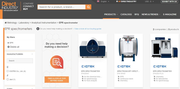
Check out online marketplaces that promote scientific instruments. These platforms allow users to find and compare prices for various scientific techniques, including ESR spectroscopy. Examples include GlobalSpec, DirectIndustry, etc. For example:

best price Electron Spin Resonance spectroscopy

best price ESR spectroscopy


3. Request multiple quotes

When contacting agencies, EPR manufacturers, or vendors, request quotes from multiple sources. Provide them with specific details about the project, including sample type, sample size, data analysis needed, and any special requirements. Comparing multiple quotes will help you find the best price. Many EPR suppliers offer free, comprehensive, customized quotes, such as CIQTEK EPR.

best price EPR spectroscopy


4. Consider geographic location

The price of EPR Spectroscopy may vary by region or country. Sometimes, purchasing EPR Spectroscopy from a country with lower labor or equipment costs can result in significant cost savings. For example, CIQTEK's self-developed EPR Spectroscopy has the world's leading EPR technology to maintain high-quality service globally for any customized service and has a very competitive price.

best price EPR spectrometer


5. Research Local Academic Institutions

Contact nearby universities, colleges, and research institutions to inquire about their ESR spectroscopy services. Academic institutions often offer advanced scientific equipment at reasonable prices, especially for researchers and students.


6. Collaborate with researchers

Consider collaborating with researchers who have access to ESR spectroscopy equipment. Many researchers are open to collaboration or offer their services at a discounted rate, especially if your project aligns with their interests. Attend scientific conferences, workshops, or networking events to connect with the relevant research community.


Keep in mind that while cost is an important factor, ensuring that the service provider delivers reliable, accurate results is also critical. Evaluate their technical expertise, reputation, and experience before deciding.

How To Choose An ESR Spectroscopy For Your Research?

There are many factors to consider when selecting an Electron Paramagnetic Resonance (EPR) spectroscopy for your research.

Some of the key points are listed below:


Frequency Range: Determine the frequency range needed for your study. EPR spectroscopy is available in different frequency ranges, such as X-band, Q-band, and W-band. The choice depends on the type of sample you are studying and the level of spectral resolution required.


Magnetic Field Strength: Consider the magnetic field strength required for your experiment. EPR spectroscopy operates at different magnetic field strengths, such as 0.35 T, 1.2 T, or higher. Higher field strengths provide better spectral resolution but may require specialized equipment and cost more.


Sample Size and Compatibility: Evaluate sample size and compatibility requirements. Some EPR spectroscopy is designed for small sample sizes, while others can accommodate larger samples or even solid-state systems. Make sure the spectroscopy you choose will meet your specific sample requirements.


Instrument Sensitivity and Signal-to-Noise Ratio: Evaluate the sensitivity and signal-to-noise ratio (SNR) of your EPR spectroscopy. Higher sensitivity and SNR will detect weaker signals and improve the overall quality of the measurement. Consider the application requirements and the sensitivity needed for the sample.


Modulation capability: Checks the modulation capability of the EPR spectroscopy. Modulation techniques such as continuous wave (CW), pulse, echo, or Fourier transform (FT) are critical in different experimental setups. Ensure that the spectroscopy supports the modulation techniques required for your study.


Additional features and accessories: Consider any additional features or accessories that may be required for the experimental setup. This may include a temperature control system, variable angle goniometer, cryostat, or specialized resonator. Evaluate whether the spectroscopy has the required flexibility and compatibility with these accessories.


Cost and Availability: Finally, consider the cost and availability of the EPR spectroscopy. The cost and availability of the various models offered by different manufacturers vary. Ensure that the EPR spectroscopy you choose fits your budget and that the manufacturer offers reliable support and service.


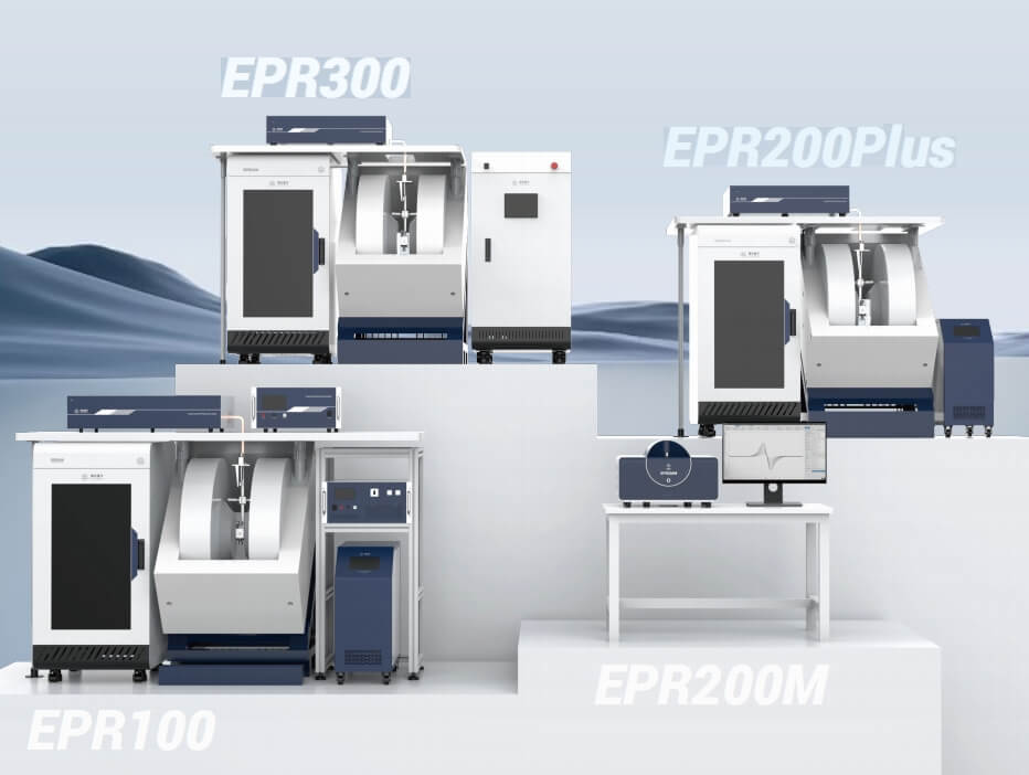
CIQTEK offers different specialized custom EPR spectroscopy solutions for different customer needs.

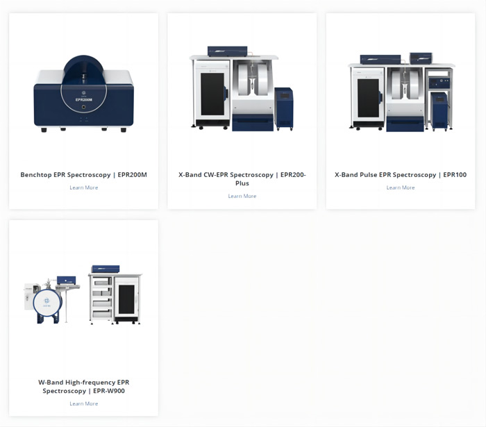
From benchtop EPR to floor-stand EPR spectroscopy, X-band to W-band EPR spectroscopy, pulsed and continuous wave EPR spectroscopy, at affordable prices and world-leading quality.

CIQTEK EPR spectroscopy should be the first choice for EPR users.

CIQTEK Scanning Electron Microscope Competitive Advantage Analysis Report

CIQTEK Scanning Electron Microscope (SEM) Competitive Advantage Analysis Report in terms of price, quality, and service:


Best Price:

CIQTEK SEM is competitively compared to other similar products on the market. The company offers a range of different models and specifications to meet the needs of different customers. By offering affordable options, CIQTEK appeals to customers looking for cost-effective solutions for their SEM microscopy needs.


High Quality:

CIQTEK is known for delivering high-quality scanning electron microscopes. The company uses advanced technology and ensures rigorous quality control in the manufacturing process. CIQTEK SEMs are designed to provide accurate and precise imaging, offer high resolution, and enable various imaging modes and analytical capabilities. The company's commitment to quality ensures that customers receive reliable and accurate results from their SEMs.


Great Service:

CIQTEK places great emphasis on customer service and support. The company has a dedicated team of trained professionals who provide technical assistance, installation support, and training to customers. CIQTEK offers prompt and efficient customer support to address any queries or issues that may arise during the lifetime of the SEM. This comprehensive support system enhances the overall customer experience and ensures that customers receive satisfactory after-sales service.


In summary, CIQTEK SEM has a competitive edge in the market due to its affordable pricing, high-quality imaging capabilities, and comprehensive customer service. These factors contribute to the company's ability to attract and retain customers in the increasingly competitive field of scanning electron microscopy.

Scanning Electron Microscope Manufacturers In South Korea

There are several scanning electron microscope manufacturers in Korea.

The following are some of the famous companies:


JEOL LIMITED: JEOL is a manufacturer of scientific instruments and equipment, including scanning electron microscopes. They offer a range of scanning electron microscope models with different specifications and applications.


Hitachi High-Tech Korea Co., Ltd: Hitachi High-Tech Korea is a subsidiary of Hitachi High-Tech Corporation, a global technology company. They offer a wide range of scientific instruments for research and industrial applications, including scanning electron microscopes.


Samsung Electronics Co. Ltd: Samsung Electronics is a leading South Korean multinational company that produces a wide range of products including consumer electronics and industrial equipment. They also produce advanced electron microscopes, including scanning electron microscopes.


S-Connect Co., Ltd: S-Connect is a South Korean company specializing in the development and manufacture of scientific instruments. They offer customized scanning electron microscopes for a variety of research and industrial applications.


TESCAN Korea Co., Ltd.: TESCAN is a global supplier of scientific instruments including scanning electron microscopes. With a branch office in Korea, the company provides SEM solutions for materials science, life science, and other fields.


CIQTEK Co., Ltd.: CIQTEK is a global manufacturer of high-value scientific instruments including Scanning Electron Microscope (SEM).

CIQTEK's SEM products offer advanced microscope technology and innovative features that provide high-resolution surface topography and compositional analysis. 

In addition, CIQTEK provides SEM-related technical support and training services to help users realize the full potential of SEM. They have a professional team with extensive experience and knowledge to provide consulting and solutions.

Check the CIQTEK SEM microscope here.


Research each company's products, specifications, and customer reviews to find the best SEM for specific needs.

A Key Factor G-Value In EPR Spectroscopy

2024-06-22

The g-value plays a crucial role in EPR (Electron Paramagnetic Resonance) spectroscopy in understanding the electronic structure and magnetic properties of paramagnetic substances. Today, we will talk about the key factor in EPR spectroscopy: the g-value (g-factor).


The g-value is a dimensionless quantity that represents a constant of proportionality between the magnetic field and the energy difference between the energy levels in the system. The g-value can be obtained by measuring the resonance frequency of a paramagnetic substance absorbing electromagnetic radiation in the presence of a magnetic field. It represents the extent to which the electron spin interacts with the external magnetic field. The g value is commonly used to determine important characteristics of paramagnetic substances, such as the number of unpaired electrons and their orbital angular momentum.


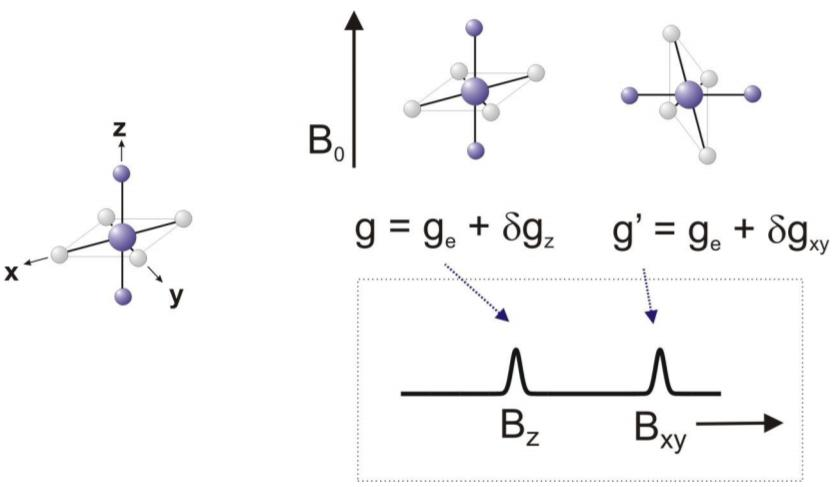
For a free electron, the g value is a constant of 2.0023, derived from fundamental physical properties. However, in more complex systems, such as transition metal complexes or organic radicals, the g value can deviate from this standard value. The deviation arises due to various factors, including spin-orbit coupling and hyperfine interactions with nearby nuclei. These interactions introduce additional energy levels and modify the electron's behavior in the magnetic field, leading to different g values.


By analyzing the g value in EPR spectroscopy, scientists can gain insights into the molecular structure and chemical environment of the paramagnetic species under investigation. A change in the g value can reveal the presence of different ligands or coordination environments around a metal ion, providing valuable information about the electronic configuration and coordination chemistry.

g value in epr spectroscopy

In addition, the g-value can be used to study the dynamics of a system. For example, it can provide information about the rate of electron transfer or spin relaxation processes, revealing the kinetics and mechanisms involved in chemical reactions or biological processes.


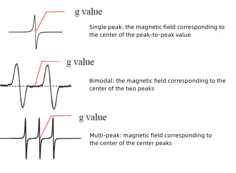
A common method of measuring the g-value is using EPR spectroscopy on a sample with a known g-value. This reference sample can be a compound with a well-characterized g-value or a spin probe, such as a stable organic radical. By comparing the position and shape of the resonance signals of the reference sample and the sample of interest, the g-value of the unknown species can be calculated.


Another approach involves calculating the g value from the Zeeman effect. The Zeeman effect describes the splitting of energy levels in a magnetic field. By measuring the magnetic field strength required to observe resonance at different frequencies, the g value can be derived using the equation g = hν/μBΔB, where h is Planck's constant, ν is the microwave frequency, μB is the Bohr magneton, and ΔB is the magnetic field difference between the resonance peaks.


In addition, the g value can be determined by analyzing the linewidth of the EPR signal. Since the g value depends on the orientation of the unpaired electrons in the magnetic field, any interaction or fluctuation in the environment will broaden the EPR signal. The g-value can be estimated by measuring the linewidth and analyzing its dependence on the magnetic field strength.


It is worth noting that the g value is not always a fixed constant. In some cases, it may vary due to factors such as electron-electron interactions or electrons leaving the domain. These variations can indicate the chemical or physical properties of the paramagnetic substance.

g value in epr spectroscopy

In summary, the g-value in EPR spectroscopy is an important parameter that helps characterize paramagnetic substances. By determining the g-value, researchers can gain valuable insights into the electronic structure and properties of paramagnetic compounds, contributing to understanding a wide range of scientific disciplines, including chemistry, physics, and biology.


Click here to read more application notes on EPR spectroscopy.

Electron Paramagnetic Resonance Spectroscopy Applications In Free Radical Detection

2024-06-22

What are free radicals?


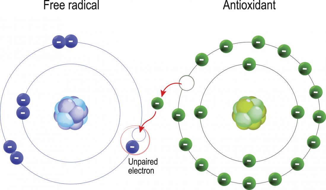
Free radicals are highly reactive molecules or atoms that play a crucial role in various chemical and biological processes. Understanding their behavior and detecting their presence is essential to studying their involvement with disease progression, environmental pollution, and other biological and chemical systems.

Image via the Internet

Image via the Internet

Principles of EPR Spectroscopy

EPR spectroscopy utilizes the magnetic properties of unpaired electrons in paramagnetic substances. When samples containing such substances are subjected to magnetic fields and electromagnetic radiation, electron spin transitions occur, resulting in the absorption or emission of energy. By measuring the occurrence of these transitions, valuable information about the paramagnetic substance and its environment can be obtained.


Application of EPR Spectroscopy in Free Radical Detection

Electron Paramagnetic Resonance (EPR) spectroscopy, also known as Electron Spin Resonance (ESR), is a powerful analytical technique used to study paramagnetic species, including free radicals. Free radicals play a crucial role in various chemical and biological processes, and their detection and understanding are essential for a range of applications. Since free radicals have unpaired electrons, they can be detected and characterized using EPR spectroscopy. This report explores the application of EPR spectroscopy in free radical detection.


1. Chemical Reactions

EPR spectroscopy provides valuable insights into the kinetics and mechanisms of free radical reactions in chemical systems. By monitoring changes in the EPR spectra during a reaction, researchers can determine the formation and consumption rates of free radicals, elucidate reaction pathways, and assess the effectiveness of radical scavengers or inhibitors.


2. Biological Systems

In biological systems, free radicals are involved in several physiological and pathological processes. EPR spectroscopy can be employed to study the generation, localization, and reactivity of free radicals in living organisms. By using spin traps or spin probes that selectively interact with specific radicals, researchers can gain insights into oxidative stress, inflammation, and disease progression.


3. Material Science

Free radicals can be formed and trapped in materials during their synthesis or under specific conditions. EPR spectroscopy allows for the detection and characterization of these trapped radicals, enabling researchers to explore their effects on the material's properties. This information contributes to the development of materials with improved performance and stability.


Conclusions

EPR spectroscopy has proven to be a valuable tool in the field of free radical detection, with applications spanning a variety of fields, including chemical reactions, biological systems, and materials science. Through the use of EPR spectroscopy, researchers can gain insight into the nature, behavior, and effects of free radicals, and thus better understand the role of free radicals in a wide range of processes.

CIQTEK has been deeply engaged in the field of EPR technology for many years and has independently developed a variety of EPR spectrometers for different research purposes, including benchtop EPR, floor-standing EPR, continuous wave EPR, pulsed EPR, etc. 

Zowell Double -deep Reach Forklift Empowers Overseas Auto Parts Factories to Realize Efficient and Intensive Storage

2024-06-20

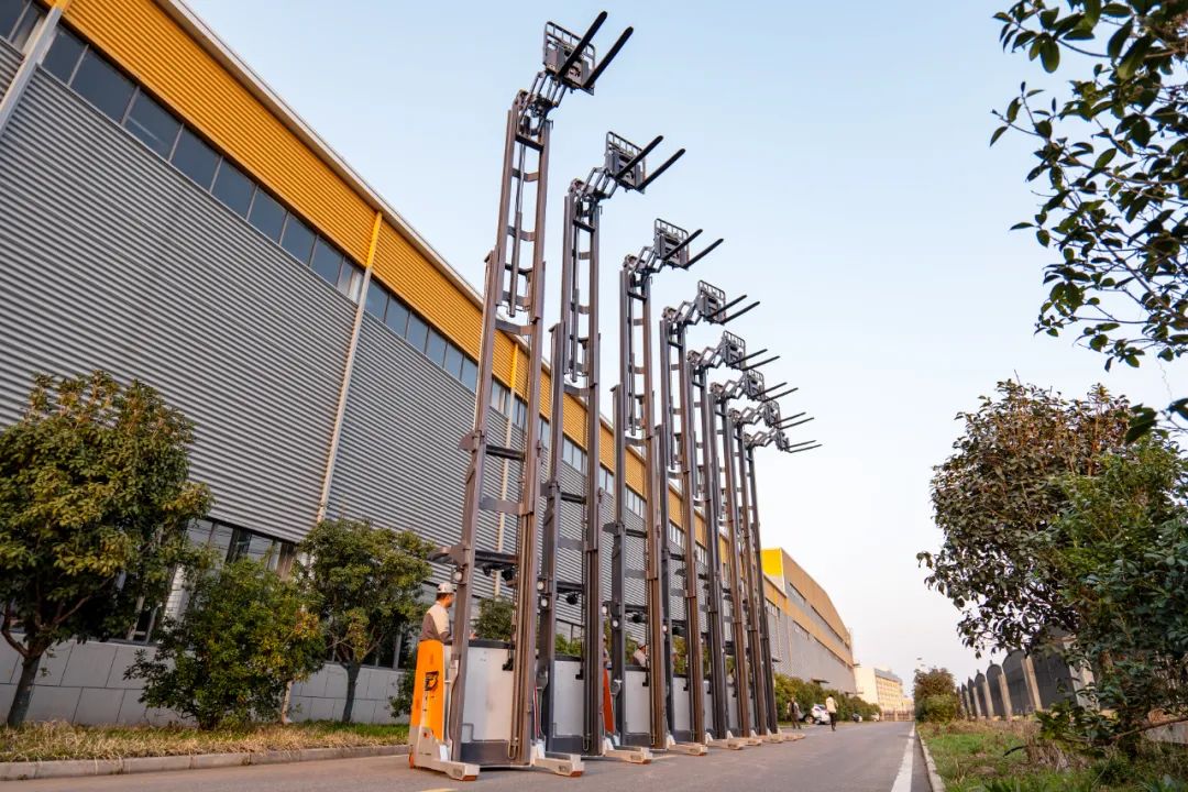
The manufacturing floor empowered by automation technology gives us a stronger and more reliable order processing capability, enabling us to deliver at an unprecedented speed. After passing the strict quality inspection, 8 sets of double-deep reach forklifts embarked on their journey to the sea.


Project Background

This large-scale procurement customer is an overseas auto parts factory, specializing in the research and development, production and sales of parts in the field of new energy vehicles, which is a leading level in the industry.

As the new energy vehicle industry is in a period of rapid growth, driving the continuous expansion of the auto parts industry. The enterprise continues to obtain a large number of orders, the urgent need for logistics upgrades, hoping to improve customer satisfaction and enhance the company's products in the global market share by virtue of advanced order processing capabilities.

The popularity of Zowell brand in the international market has been increasing. After taking the initiative to contact and learn about our successful cases of serving other enterprises and more flexible customized services, the customer firmly chose JOYAL to realize the upgrading of the internal logistics of the enterprise!


Existing Problems

1. With the expansion of the business scale and the increase of SKUs, the pressure of the enterprise's warehousing has been highlighted. However, the cost of expanding the warehouse is high, so how to realize the improvement of the storage volume rate in the existing warehouse has become a new challenge.

2. From the raw material warehouse to the docking production line, the finished product transfer into the warehouse, each process must be closely linked, however, the current logistics handling equipment of the enterprise is inefficient, the internal transfer cycle is long, and the delivery of orders is slow.

3. Heavy-duty, palletized auto parts have certain risks when handling, the enterprise attaches great importance to the safety of employees and the factory, and hopes to have a breakthrough in safety and security.

Zowell Solution

Part.1

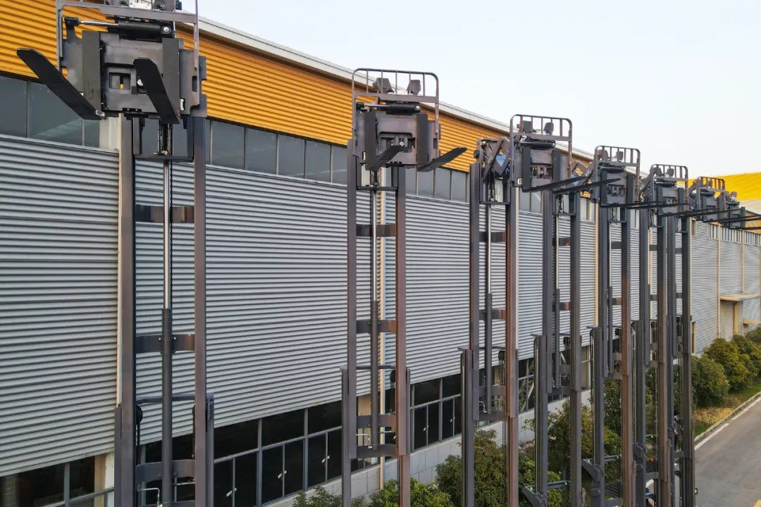
The enterprise Zowells suggestion to reconstruct the warehouse space, and used 11m high double-extension racks to cooperate with our double-deep reach trucks to carry out the warehouse operation. The space on the upper floor of the warehouse was fully released, the overall access area of the warehouse was greatly reduced, and the warehouse realized a nearly 140% increase in storage space while the land cost remained unchanged.

Part.2

Equipped with an 8.0 kw traction motor, Zowell double-deep reach trucks can reach a maximum speed of 10km per hour and start-stop driving without skidding, with excellent driving ability, which can maximize the efficiency of cargo transfer between warehouse-connection point-within warehouse under safe conditions.

Part.3

Zowell attaches great importance to the safety and durability of the product, the German imported channel steel door frame and solid roof rack can allow the driver to operate in the vehicle body protection completely within the scope of the protection, by a full range of protection.

Part.4

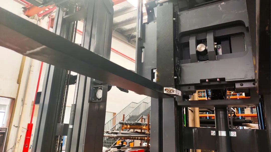
Double-deep reach truck can load 1.5T of goods, in the fork is also equipped with high-definition wireless camera, no matter empty/full load, high/low position, the driver can "zero distance" clearly observe the position of the warehouse, the pallet position, to avoid misoperation to produce a collision, jeopardizing the safety of people and property.

Tags